Thighbone (Femur) Fractures In Children
The thighbone (femur) is the largest and strongest bone in the body. It can break when a child experiences a sudden forceful impact.
Cause
Statistics
The most common cause of thighbone fractures in infants under 1 year old is child abuse. Child abuse is also a leading cause of thighbone fracture in children between the ages of 1 and 4 years, but the incidence is much less in this age group.
In adolescents, motor vehicle accidents (either in cars, bicycles, or as a pedestrian) are responsible for the vast majority of femoral shaft fractures.
Risk
Events with the highest risk for pediatric femur fractures include:
- Falling hard on the playground
- Taking a hit in contact sports
- Being in a motor vehicle accident
- Child abuse
Types of Femur Fractures (Classification)
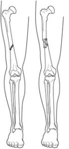
Femur fractures vary greatly. The pieces of bone may be aligned correctly (straight) or out of alignment (displaced), and the fracture may be closed (skin intact) or open (bone piercing through the skin). An open fracture is rare.

Specifically, thighbone fractures are classified depending on:
- Location of fracture on the bone (proximal, middle, or distal third of the bone shaft)
- Shape of the fractured ends — bones can break all kinds of ways, such as straight across (transverse), angled (oblique), or spiraled (spiral)
- Position of the fractured edges (angulated or displaced)
- Number of fractured parts
- Two parts
- Several fractured parts (comminuted)
Symptoms
A thighbone fracture is a serious injury. It may be obvious that the thighbone is fractured because:
- Your child has severe pain
- The thigh is noticeably swollen or deformed
- Your child is unable to stand or walk, and/or
- There is a limited range of motion of the hip or knee allowed by the child because of pain.
Take your child to the emergency room right away if you think he or she has a broken thighbone.
Doctor Examination
It is important that the doctor know exactly how the injury occurred. Tell the doctor if your child had any disease or other trauma before it happened.
The doctor will give your child pain relief medication and carefully examine the leg, including the hip and knee. A child with a thighbone fracture should always be evaluated for other serious injuries.
Imaging Tests
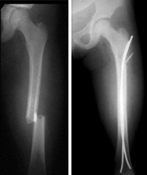
Your orthopaedic doctor will need x-rays to see what the broken bone looks like (refer to “Classification”). Your child’s healthy leg may also be x-rayed for comparison.
The orthopaedic doctor will also check the x-ray for any damage to the growth area (growth plate) near the end of the femur. This is the part that enables the child’s bone to grow. If needed, surgery may help to restore the growth plate’s function, and regular x-rays may be taken for many months to track the bone’s growth.
Treatment
To treat a child’s thighbone fracture, the pieces of bone are realigned and held in place for healing. Treatment depends on many factors, such as your child’s age and weight, the type of fracture, how the injury happened, and whether the broken bone pierced the skin.
Nonsurgical Treatment

In some thighbone fractures, the doctor may be able to manipulate the broken bones back into place without an operation (closed reduction). In a baby under 6 months old, a brace (called a Pavlik Harness) may be able to hold the broken bone still enough for successful healing.
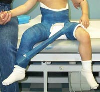
Spica casting. In children between 7 months and 5 years old, a spica cast is often applied to keep the fractured pieces in correct position until the bone is healed.
There are different types of spica casts, but, in general, a spica cast begins at the chest and extends all the way down the fractured leg. The cast may also extend down the uninjured leg, or stop at the knee or hip. Your doctor will decide which type of spica cast is most effective for treating your child’s fracture.
Your doctor will sedate your child for the closed reduction, and apply a spica cast immediately (or within 24 hours of hospitalization) to keep the fractured pieces in correct position until healing occurs.

When a bone breaks and is displaced, the pieces often overlap and shorten the normal length of the bone. Because children’s bones grow quickly, your doctor may not need to manipulate the pieces back into perfect alignment. While in the cast, the bones will grow and heal back into a more normal shape.
In general, for the best results, the broken pieces should not overlap more than 2 cm when in the cast. The growth of the thighbone may be temporarily increased by the trauma. The mild shortening from the overlap will resolve.
Traction. If the shortening of the bones is too much (more than 3 cm) or if the bone is too crooked in the cast, it may be helpful to put the leg in a weight and counterweight system (traction) to make sure the bones are properly realigned.
Surgical Treatment
Doctors generally agree that displaced femur fractures that have shortened more than 3 cm are not acceptable and require treatment to correct at least a portion of the shortening.

In some more complicated injuries, the doctor may need to surgically realign the bone and use an implant to stabilize the fracture.
Doctors are treating pediatric thighbone fractures more often with surgery than in previous years due to the benefits that have been recognized. These include earlier mobilization, faster rehabilitation, and shorter time spent in the hospital.
In children between 6 and 10 years old, flexible intramedullary (inside the bone) nails are often used to stabilize the fracture. Over the past decade, this treatment method has gained great acceptance.
Occasionally, the broken bone has too many pieces and cannot be treated successfully with flexible nails. Other options that can lead to successful outcomes in this situation include:
- A plate with screws that “bridges” the fractured segments
- An external fixator — this is often used if there has been a large open injury to the skin and muscles
- Prolonged traction with a pin temporarily placed into the thighbone

As the child nears the teenage years (11 years to skeletal maturity), the most common treatment choices include either flexible intramedullary nails or a rigid locked intramedullary nail. The rigid nail is particularly useful when the fracture is unstable. Both types of nails allow for the child to begin walking immediately.

Long-Term Outcomes
Generally, children who sustain a thighbone fracture will heal well, regain normal function, and have legs that are equal in length. The intramedullary nails may need to be removed following healing if they cause irritation of the skin and tissues underneath.
Occasionally, children will require further treatment, either early on or in subsequent years, if they have a significant difference in the length of the legs, unacceptable angulation of the healed bone, abnormal rotation of the healed bone, infection, or (rarely) if a thighbone fracture persists (nonunion).
These problems can nearly always be resolved with further treatment.
For More Information
If you found this article helpful, you may also be interested in Internal Fixation for Fractures.
In order to assist doctors in the treatment of thighbone fractures, the American Academy of Orthopaedic Surgeons has done research to provide some useful guidelines. These are recommendations only and may not apply to each and every individual case. For more information: AAOS Clinical Practice Guideline: Treatment of Pediatric Diaphyseal Femur Fractures

Reviewed by members of POSNA (Pediatric Orthopaedic Society of North America)
